Figure 1. Proliferative diabetic retinopathy epiretinal membranes. A: Immunohistochemical staining of panendothelial cell marker CD34 shows blood vessels positive for CD34 (arrows; original
magnification 40×). Immunohistochemical staining of high-mobility group box −1 (HMGB1). Low power (B; original magnification 40×) and high power (C; original magnification 100×) showing vascular endothelial (arrows) and stromal (arrowheads) cells expressing strong immunoreactivity
to HMGB1. Immunohistochemical staining of receptor for advanced glycation end products (RAGE). Low-power (D; original magnification 40×) and high-power (E; original magnification 100×) showing vascular endothelial (arrows) and stromal (arrowheads) cells expressing strong immunoreactivity
to RAGE. Immunohistochemical staining of osteopontin (OPN) showing vascular endothelial cells (arrows; F) and stromal cells (arrowheads; G) expressing strong immunoreactivity to OPN (original magnification 100×). Immunohistochemical staining of early growth response-1
showing immunoreactivity in vascular endothelial (arrows) and stromal (arrowheads) cells (H; original magnification 100×).
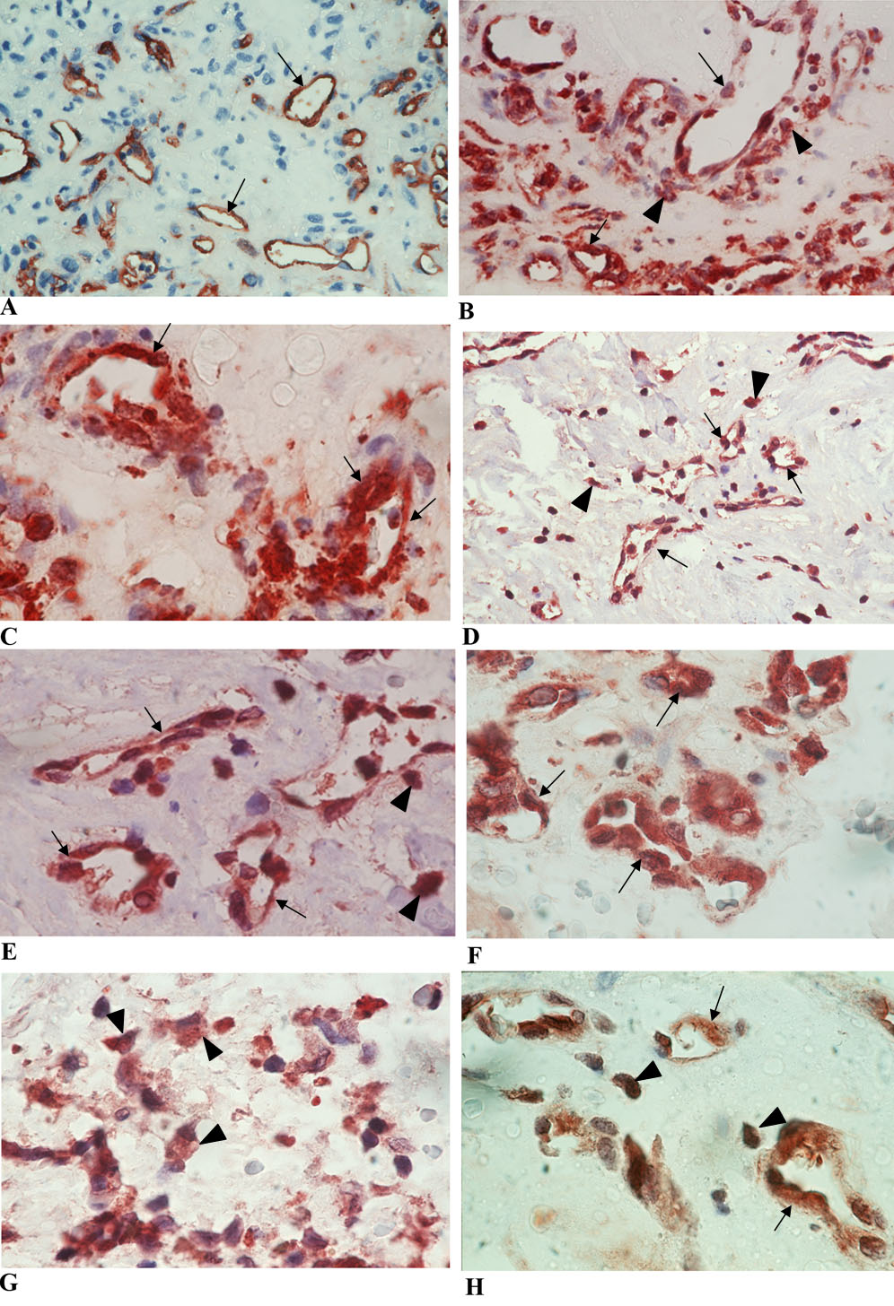
 Figure 1 of
Abu El-Asrar, Mol Vis 2011; 17:508-518.
Figure 1 of
Abu El-Asrar, Mol Vis 2011; 17:508-518.